
Echocardiogram Test
Advanced echo scans for precise, real-time heart evaluation
Echocardiogram (2D, 3D & Stress Echo)
Overview
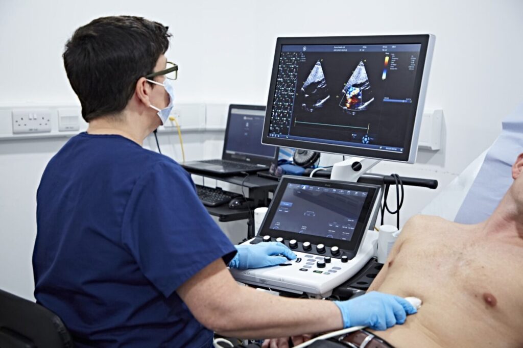
An Echocardiogram—commonly called an “echo”—is a sophisticated ultrasound imaging test that creates detailed, moving pictures of your heart. This safe, painless, non-invasive procedure allows your cardiologist to see your heart’s structure, watch how it pumps blood, evaluate valve function, and assess overall cardiac performance in real-time.
At Good Heart Clinic, we utilize state-of-the-art echocardiography technology including advanced 2D imaging, cutting-edge 3D visualization, and specialized stress echocardiography to provide the most comprehensive heart assessment available. Whether you’re experiencing symptoms, have been diagnosed with heart disease, or need routine monitoring, an echocardiogram provides critical visual information that guides accurate diagnosis and optimal treatment planning.
Echocardiogram in Pune | 2D Echo Test
If you are looking for a reliable echocardiogram in Pune, Good Heart Clinic offers advanced 2D Echo testing to evaluate heart structure and function with precision.A 2D Echo test in Pune uses ultrasound waves to create real-time images of the heart, helping detect valve disorders, heart muscle weakness, and other cardiac conditions.
What is an Echocardiogram?
An echocardiogram is a diagnostic imaging test that uses high-frequency sound waves (ultrasound) to create detailed, real-time images of your heart’s structure and function. Just like ultrasound imaging used during pregnancy to see a baby, an echocardiogram uses the same safe technology to visualize your heart beating, blood flowing through chambers and valves, and the heart muscle contracting.
How It Works
The echocardiogram procedure involves:
- A transducer (ultrasound probe) is placed on your chest, coated with a special gel
- High-frequency sound waves are emitted from the transducer
- Sound waves bounce off your heart structures and return to the probe
- A computer processes these echoes and converts them into moving images
- Real-time video of your beating heart appears on a monitor
Types of Echocardiogram Test
We offers several types of echocardiography, each designed for specific diagnostic needs:
Transthoracic Echocardiogram (TTE) - Standard 2D Echo
What it involves: The most common type of echo where a transducer is placed on your chest wall to obtain images through your chest (transthoracic means "across the chest").
Three-Dimensional Echocardiogram (3D Echo)
What it involves: Advanced echocardiography that creates three-dimensional images of your heart, providing volumetric visualization that's more accurate than traditional 2D imaging.
Stress Echocardiogram (Stress Echo)
Combines standard echocardiography with exercise stress testing or medication-induced stress to evaluate how your heart functions under increased workload.
Symptoms
Chest Pain
Pressure, tightness, or squeezing in the chest as well as Chest pain triggered by exertion
Shortness of Breath
Difficulty breathing with exertion or at rest. Breathlessness that's gotten progressively worse
Heart Murmur
Your doctor heard an abnormal sound while listening to your heart with a stethoscope
Swelling (Edema)
Swelling in legs, ankles, or feet. Also, Sudden weight gain from fluid retention
Palpitations
Heart racing, pounding, or fluttering as well as Sensation of irregular heartbeat
Dizziness / Fainting
Feeling lightheaded, especially with exertion. Dizziness that occurs with palpitations
Fatigue & Weakness
Unusual, persistent tiredness. Inability to perform activities you previously could do
Heart Attack Symptoms
Current or recent chest pain concerning for heart attack.
Benefits
Completely Safe and Non-Invasive
Accurate Assessment of Heart Function
Unmatched Valve Assessment
Comprehensive Cardiac Evaluation
Preparations
Preparation for an echocardiogram varies depending on the type of echo you’re having. Here’s what you need to know:
For Standard Transthoracic Echocardiogram (2D/3D Echo)
Eating and Drinking:
Clothing
For Stress Echocardiogram
More preparation needed due to exercise component:
Fasting:
- Do not eat or drink for 3-4 hours before your test
- Small sips of water are usually permitted
- Avoid caffeine (coffee, tea, energy drinks, chocolate) for at least 12 hours before
Clothing:
- Wear athletic/exercise clothing and comfortable walking shoes
- Men: comfortable shorts and t-shirt, athletic shoes
- Women: sports bra, comfortable shorts/pants, t-shirt, athletic shoes
- NO sandals, dress shoes, or flip-flop
What NOT to Do
For Standard/3D Echo:
For Stress Echo:
Procedure
Step 1: Preparation (5-10 minutes)
- You’ll lie on your left side on the examination table
- Pillow provided for your head
- Your left arm positioned above your head or comfortably out of the way
- Blanket provided for warmth and modesty (rooms kept cool for equipment)
Step 2: The Echocardiogram Imaging (30-45 minutes)
Gel Application
- Warm ultrasound gel applied to your chest
- Gel allows the ultrasound probe to glide smoothly and ensures good contact
- May feel slightly cool or warm depending on room temperature
The Ultrasound Probe (Transducer)
- A smooth, hand-held device about the size of a computer mouse
- Technician (cardiac sonographer) will place probe on various positions on your chest
- Firm pressure is applied – this is normal and necessary to get clear images through ribs and tissue
- Pressure may feel uncomfortable but shouldn’t be painful
- Speak up if pressure becomes too uncomfortable
The technician will obtain images from several positions:
Parasternal Window (left chest):
- Probe placed to the left of your breastbone
- Views your heart’s long and short axes
- Visualizes left ventricle, right ventricle, valves
Apical Window (under left breast):
- Probe positioned at the point where you can feel your heartbeat (apex)
- Shows all four chambers
- Critical for measuring ejection fraction
- May require lifting breast tissue (women) for proper positioning
Subcostal Window (upper abdomen):
- Probe placed just below your ribcage
- You may be asked to take a deep breath and hold it
- Provides views of heart from below
- Especially useful in patients with lung disease
Suprasternal Window (above collarbone):
- Probe placed in the notch above your sternum
- Visualizes the aorta
- May feel unusual but not painful
Step 3: Completion and Cleanup (5 minutes)
Finishing Up
- Technician will remove ECG electrodes
- You’ll wipe off ultrasound gel with provided towels (it’s water-based and non-staining)
- Some gel may remain – easily washes off with soap and water later
- You’re free to get dressed
Immediate Post-Echo:
- No recovery time needed
- No restrictions on activities
- You can eat, drink, drive, work immediately
- No side effects or aftereffects
Frequently Asked Question (FAQs)
An ECG provides a visual representation of your heart rhythm, heart rate, and electrical patterns. It can quickly identify arrhythmias, ischemia, heart block, electrolyte imbalances, and other abnormalities. It is commonly used alongside TMT, 2D Echo, and Holter Monitoring for a complete cardiac evaluation.
Yes, an ECG is one of the most effective tests to detect ongoing or previous heart attacks, changes in heart muscle, and abnormalities in blood flow. If the ECG is abnormal, your cardiologist may recommend cardiac enzymes, 2D Echo, or coronary angiography for further clarity.
A standard resting ECG takes 5–10 minutes, making it one of the fastest and most convenient cardiac tests. Reports are generated immediately and interpreted by experienced cardiologists for instant results.
An ECG can detect electrical abnormalities and signs of reduced blood flow, but it does not confirm the severity of blockages. For detailed assessment, doctors may advise Treadmill Stress Test (TMT), CT Coronary Angiography, or Coronary Angiogram.
Yes, ECG is a completely safe, non-invasive, and radiation-free test suitable for all age groups, including seniors and patients with chronic conditions. It is often used as a routine cardiac check-up for high-risk individuals.