
Intravascular Imaging
Overview
Intravascular Imaging is one of the most advanced cardiac diagnostic tools used to visualize the inside of coronary arteries with exceptional clarity. Traditional angiography shows only the outline of blood vessels, but intravascular imaging provides high-quality, detailed images of artery walls, blockages, plaque composition, and stent expansion. This helps cardiologists perform more accurate, safer, and highly successful treatments—especially in complex cases of coronary artery disease.
At Good Heart Clinic, intravascular imaging is used during angioplasty, stenting, and rotablation procedures to ensure precise diagnosis and optimal results. It helps your doctor understand the exact severity of blockages, identify calcium levels, assess vessel size, and confirm whether a stent has opened properly. With this technology, cardiologists avoid guesswork and make decisions based on real-time, microscopic-level information.
Intravascular Imaging in Pune
If you are looking for advanced intravascular imaging in Pune, Good Heart Clinic provides expert evaluation using cutting-edge coronary imaging techniques such as IVUS and OCT to ensure accurate diagnosis and optimal treatment planning.
Intravascular imaging in Pune enhances the precision of coronary angioplasty and stent placement by providing detailed images from inside the artery.
What is a Intravascular Imaging ?
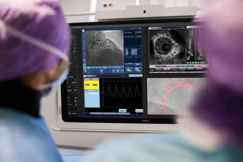
Intravascular Imaging is a specialized technique that uses miniature imaging devices placed inside the coronary artery to obtain detailed visual information about the vessel’s internal structure.
There are two main types:
IVUS (Intravascular Ultrasound)
A tiny ultrasound probe is attached to a catheter and inserted into the artery. It produces cross-sectional images of the vessel, showing plaque thickness, calcium deposits, and the exact artery size.
OCT (Optical Coherence Tomography)
OCT uses light waves to produce extremely high-resolution images, almost like a microscope. It helps visualize fine details like stent struts, micro-calcification, and plaque composition.
Time Taken for the Procedure
Intravascular imaging is quick, safe, and usually performed during angioplasty or stenting.
The procedure
Complete angioplasty with intravascular imaging takes 45 minutes to 1.5 hours, depending on complexity.
Recovery and observation
Post-procedure observation lasts 4 to 8 hours, depending on whether the wrist or groin was used.
Total hospital time
Most patients return home the same day unless prolonged monitoring is needed.
Symptoms
Persistent Chest Pain
When angiography appears normal but symptoms continue, intravascular imaging helps detect hidden blockages or micro-plaque.
Calcified Coronary Blockages
If blockages are heavily calcified or difficult to assess, IVUS or OCT helps identify plaque type and guide treatment.
Recurrent Symptoms After Stenting
If a patient develops chest pain months or years after stent placement, imaging can check for stent narrowing or incomplete expansion.
Heart attack symptoms
If you've experienced or are experiencing severe chest pain, pain radiating to your arm or jaw, sweating, nausea, or shortness of breath
Angiography Results
When angiography does not clearly show the blockage severity, intravascular imaging provides accurate measurements.
Complex Multivessel Disease
Intravascular imaging helps plan the right stent size and length for multiple blockages.
High-Risk Angioplasty
Imaging ensures precise plaque modification and optimal stent deployment.
Stress-Triggered Discomfort
If anxiety or emotional strain brings on chest or heart-related sensations.
Benefits
Accurate Assessment of Blockages
Improves Angioplasty Success
Detects Hidden Heart Problems
Enhances Long-Term Heart Health
Preparations
Although intravascular imaging is often done during angioplasty, proper preparation helps ensure a safe and smooth experience.
Before the Day of Your Procedure:
On the Day of Your Procedure:
What Does the Procedure Involve?
Intravascular imaging is performed in the cath lab, usually alongside an angioplasty or diagnostic coronary procedure.
Here is what typically happens:
- The wrist or groin area is cleaned and numbed with local anesthesia.
- A catheter is inserted and guided toward the coronary artery.
- An IVUS or OCT catheter is inserted through the guiding catheter into the artery.
- Real-time images of the artery are taken as the imaging catheter moves through the blockage site.
- The doctor evaluates plaque thickness, calcium, vessel shape, and artery size.
- Based on the findings, balloon angioplasty, stent placement, or plaque modification is performed.
- Imaging is repeated after stenting to confirm proper stent expansion.
- The catheter is removed and a closure band or device is applied.
The entire imaging process is safe, painless, and highly precise.
What Follows the Procedure?
After intravascular imaging and angioplasty, patients are taken to the recovery area for monitoring.
First Week Recovery
- Rest adequately and avoid strenuous activities.
- Keep the wrist or groin site clean and dry.
- Avoid raising heavy objects or bending the wrist forcefully.
- Light walking is encouraged to improve circulation.
- Take all prescribed medications daily.
- Follow a heart-healthy diet and stay hydrated.
- Attend follow-up appointments for stent and heart function evaluation.
Warning Signs to Watch For
Seek immediate medical help if you notice:
- Persistent chest pain
- Severe shortness of breath
- Bleeding or swelling at the catheter site
- Fever or signs of infection
- Rapid or irregular heartbeat
- Dizziness or fainting
- Severe arm or leg discomfort depending on the catheter site
Early intervention helps prevent complications.
Frequently Asked Question (FAQs)
No. Intravascular imaging is not painful because it is performed under local anesthesia during an angioplasty procedure. Patients may feel slight pressure at the wrist or groin insertion site but no significant discomfort.
Not always. It is used mainly for:
- Complex or calcified blockages
- Unclear angiography results
- Multivessel disease
- Stent-related complications
- Heart attacks requiring detailed assessment
Doctors suggest angiography when you have warning signs of heart blockage such as chest pain, breathlessness, fatigue, dizziness, or abnormal stress test results. Patients with diabetes, high BP, or high cholesterol may also need this test earlier for accurate heart disease detection.
Yes. It is a very safe and commonly performed cardiac test. Complications are extremely rare, especially when done by an experienced cardiologist. Your medical team monitors you throughout the procedure to ensure complete safety.
Yes. Coronary angiography is considered the gold standard for detecting coronary artery blockages. It shows the exact location, size, and severity of narrowing in the arteries, which helps in planning treatment like stenting or bypass surgery.